
We visited the Francis Crick Institute in London recently to attend the BioBeat17 summit, which focused on the theme ‘Re-shaping Biotech Partnering’. At the meeting, we heard Dr Lucy Collinson, Head of Electron Microscopy at the Crick, present a fascinating case study that illustrated how collaboration can drive innovation and translation within biomedical research.
By assembling a multidisciplinary team and establishing partnerships with various biotech companies, Dr Collinson managed to physically combine a fluorescent light microscope and a volume electron microscope for the first time, supported by the Crick’s scientific translation team and its distinctive ‘discovery without boundaries’ strategy. Below we uncover how this unique synergy has changed the face of imaging technology, to reveal a fresh viewpoint on disease biology.
How to solve a 'needle in a haystack' problem
In her talk, Dr Collinson explained how she and her team have been working to resolve what molecular biologists often call a 'needle in a haystack' problem. This refers to the challenge of locating a particular molecular event in space and time that is vital to understanding a disease pathway, but is exceptionally rare when you take into account the sheer number of cells you have to sift through. Such events include a single blood vessel fusion that enables a cancerous tumour to grow, or a cell becoming infected with a particular intracellular pathogen.
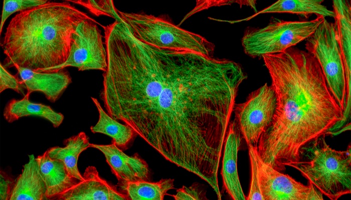
Dr Collinson explained how correlative light and electron microscopy (CLEM) is opening new avenues for the investigation into these problems. Using a spectrum of dyes and probes, fluorescence light microscopy enables us to locate molecules of interest within living cells, while high-resolution electron microscopy (EM) reveals the ultrastructure of cells and tissues. As such, combining these two methods in CLEM enables the initial location and function of the fluorescent proteins that characterize the rare ‘needle in a haystack’ event, before following this up with EM analysis to obtain crucial nano-scale information.
However, CLEM technologies have had some drawbacks in the past, including crucial differences in the sample preparation methods needed for each type of microscopy. For example, for electron microscopy, the samples must be stained with heavy metals to give contrast in the microscope, and embedded in resin to protect them from the vacuum in the microscope chamber. This makes it difficult to preserve the fluorescent markers for analysis under the microscope.
“Large microscopy companies have previously developed correlative imaging systems that have either not had good uptake in the market or have been shelved" Dr Collinson explained. “I think it’s because these companies haven’t worked with the scientists who have to use the systems and do the sample preparation".
Taking inspiration from collaboration
Dr Collinson set out to develop a new way to image rare cellular events that overcomes the limitations of existing CLEM technologies, and allows for the identification of these events via fluorescence techniques, for further examination via EM.
Firstly, she assembled a multidisciplinary team familiar with electron microscopy engineering, including physicists Dr Martin Jones and Dr Lizzy Brama, as well as neuroscientist Dr Chris Peddie. Then, she set up partnerships with small- to medium-sized biotech enterprises with expertise in microscope design and manufacture. Through this collaborative approach, Dr Collinson's team have now managed to design and develop novel integrated light and electron microscopes.
Researchers can now use this novel correlative microscopy method to image macromolecular functions within the context of cell and tissue structure for the first time. To enable this type of work, the team developed a new sample preparation protocol based on a method called resin-embedding, which preserves the fluorescence of the sample under the light microscope, while allowing it to be stained and embedded for analysis by the electron microscope in a vacuum environment.
The first tool they developed was the ‘miniLM’, which is similar in concept to fluorescence endoscopy systems used in fluorescence-guided surgery. This comprises a miniaturised fluorescent microscope that fits into a serial block-face scanning electron microscope, to enable ‘smart tracking’ of fluorescent molecules during automated serial electron image acquisition from large cell and tissue volumes.
The second tool, the ‘ultraLM’, consists of a fluorescence microscope that integrates with an ultramicrotome. This enables the ‘smart collection’ of ultrathin (50-200nm) sections of fluorescent cells or tissues from specific regions of interest for subsequent transmission electron microscopy or array tomography.
Getting the technology into the lab
Adding more fuel to the fire was the team’s plan to make the technology open source, so that the entire electron microscopy community could build similar versions in their own labs. As such, the team published the research paper in Wellcome Open Research, attended conferences to spread the word, and ran workshops to train people on how to build and use the new machines.
However, one question remains about how the conflict between open source technology versus its future commercialisation can be successfully managed. As Dr Collinson explained, “most research groups won’t have the capabilities to build it from the open source manual, so we also want to license the technology to a company for commercial delivery to those people".
Looking ahead
Future plans are already picking up steam. Dr Collinson’s talk also revealed another collaborative project to achieve even more extreme miniaturisation of the fluorescence microscope, so it can fit into new automated electron microscopes. “This will enable the automated generation of thousands of serial images overnight through whole volumes of cells and tissues, which means we can now find the needle in the three-dimensional haystack” Dr Collinson explained. “Also, with these systems we can use software that automatically follows our cells of interest and finds them for us, to speed up discovery".
It’s safe to say that Dr Collinson's presentation left us feeling quite awestruck. We were not only impressed by the scientific innovation involved, but also by the immense potential of collaboration in overcoming some of our biggest challenges in research. It got us thinking, what else can we achieve if we continue knocking down barriers and connecting multiple disciplines and sectors?
As BioBeat founder Miranda Weston-Smith said in her opening address at BioBeat17: “It’s crucial now that we dig into our partnership imagination, to accelerate the cohesion of collaborations and shape how they can be practically achieved. It’s in our hands to make real differences". We couldn’t agree with her more!
If you want to stay up to date on the latest news in science and marketing, sign up to our newsletter today.